Thursday, October 15, 2015

The Cords Not Cut After Birth Of a Child

BY CARL ZIMMER


A team of pathologists Leiden University Medical Center in the Netherlands recently carried out an experiment that might seem doomed to failure.

They collected tissue from 26 women who had died during or just after pregnancy. All of them had been carrying sons. The pathologists then stained the samples to check for Y chromosomes.

The scientists were looking for male cells in female bodies. And their search was successful.

As reported in the journal Molecular Human Reproduction recently, the researchers found male cells in every female organ they studied: brains, hearts, kidneys and others.

In the 1990s, scientists found the first clues that cells from both sons and daughters can escape from the uterus and spread through a mother’s body. They called the phenomenon fetal microchimerism, after the chimera, a monster from Greek mythology that was part lion, goat and dragon.

But fetal cells don’t just drift passively. Studies of female mice show that fetal cells that end up in their hearts develop into cardiac tissue.

“They’re becoming beating heart cells,” said Dr. J. Lee Nelson, an expert on microchimerism at the Fred Hutchinson Cancer Research Center in Seattle.

The new study suggests that women almost always acquire fetal cells each time they are pregnant.
They have been detected as early as seven weeks into a pregnancy. In later layers, the cells may disappear, but sometimes the cells settle in for a lifetime.

In a 2012 study, Dr. Nelson and her colleagues examined the brains of 59 deceased older women and found Y chromosomes in 63 percent of them. (Many studies on fetal microchimerism focus on the cells left behind by sons, because they are easier to distinguish from the cells of their mother.)

Experts now believe that microchimerism is far from rare.

“Most of us think that it’s very common, if not universal,” Dr. Nelson said. But it remains quite mysterious.

In recent years, researchers have found many clues suggesting that microchimerism can effect a woman’s health. Tumors may be loaded with fetal cells, for example, suggesting that they might help drive cancer. Yet other studies have suggested that fetal microchimerism protects women against the disease.

“In each instance of a disease, it seems like there is this paradox,” said Amy M.Boddy, a postdoctoral fellow at Arizona State University.

Fetal microchimerism has been found in a number of mammal species, including dogs, mice and cows.

It’s likely that fetal cells have been a part of maternal life for tens of millions of years.

Microchimerism is something that humans have been evolving with since before we were humans, “said Melissa Wilson Sayres, a biologist at Arizona State.

During that time, fetal cells could have evolved into more than just bystanders. Writing in the journal  in August, Dr. Boddy, Dr.Sayres and their colleagues suggested that fetal cells may produce chemicals that influence the mother’s biology, allowing fetuses to manipulate her from within.

Some cells may help maintain the health of the mother- For example, by healing wounds. But there is also an evolutionary conflict of interest between mothers and their young.

A mother’s reproductive success depends on the total number of children she raises to adulthood over the course of her life. Devoting too many resources to a single child may leave her too frail to care for later children.

If a child can somehow coax its mother to provide more resources, on the other hand, he or she may be more likely to survive to adulthood and reproduce. Fetal cells may let children manipulate their mothers to this end, Dr. Sayres and her colleagues suggest.

Fetal cells are frequently found in breast tissue, even in milk, for instance. The researchers argue that children might thrive more if their fetal cells drove up milk production.

Mothers also nurture their babies with body heat. The thyroid gland, located in the neck, acts like a thermostat, and fetal cells in the thyroid gland in theory could cause mothers to generate more heat than they would otherwise.

This biological tension might help explain how fetal microchimerism sometimes causes harm to a mother. It may simply be an occasional side effect of the cells ‘manipulations.

There are some clues that mothers, too, pull hard in this evolutionary tug of war. The immune system kicks into high gear after giving birth, possibly to clear away left-over fetal cells. This defense may pose its own risks: Women with auto immune disorders such as rheumatoid arthritis can have relapses after pregnancy.

Some straightforward experiments could put all of these ideas to the test. Scientists could look at which genes become active in fetal cells in different parts of the body, for example. They could then see how the activity of the genes influenced a mother’s physiology, such as the production of milk.

If the preliminary results hold up, Dr. Boddy and her colleagues suggest scientists should consider how fetal cells in the brain might influence women’s behavior.

“It’s the most exciting part, but it’s the part where there’s the least research at the moment,” said Athena Aktipis, a psychologist at Arizona State and an author of the Bioessays article.” There may be a role of microchimerism in postpartum mental health.”

Dr. Nelson, who was not involved in the new paper, said that is raised a lot of ideas worth pursuing. “It’s going to be interesting to see how the data coming in over the next several years stacks up,” she said.


Taken from TODAY Saturday Edition, The New York Times International Weekly, October 3, 2015
Wednesday, July 15, 2015

Food May Be Too Clean For Our Gut’s Good Health

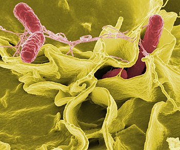
Color-enhanced scanning electron micrograph sh...
Color-enhanced scanning electron micrograph showing Salmonella typhimurium (red) invading cultured human cells (Photo credit: Wikipedia)
BY KATE MURPHY


With the recent recalls of millions of liters of ice cream as well as several tons of hummus, pine nuts, frozen vegetables and various meat products, you might think the American food supply is an unholy mess. It’s not. It’s arguably the safest in the world.

Yet despite continually improving processing methods and quality controls, the number of cases of food-borne illness has remained high since the 1990s, with the incidence of people getting sick from some pathogens increasing.

Some experts wonder if we’ve reached a point of diminishing returns in food safety-whether our food could perhaps be too clean.

Industrial food sanitation practices – along with home cooks’ antibacterial veggie washes, chlorine bleach kitchen cleaners and sterilization cycle dishwashers – kill off so-called good bacteria naturally found in foods that bolster our health. Moreover, eliminating bad or pathogenic bacteria means we may not be exposed to the small doses that could inoculate us against intestinal crises.

“No one is saying you need to eat a peck of dirt before you die to be healthy,” said Jeffrey T. Le Jeune, a professor and head of the food animal research program at Ohio State University in Wooster. “But there is a line somewhere when it comes to cleanliness. We just don’t know where it is.”

The theory that there might be such a thing as “too clean” food stems from the hygiene hypothesis, which has been gaining traction over the last decade. It holds that our modern germaphobic ways may be making us sick by harming our microbiome, which is the system made up of all the microbes-bacteria, viruses, fungi, mites-that live in and on human bodies.

A result of a diminished microbiome is an immune system that gets bored, spoiling for a fight and apt to react to harmless substances ad even attack the body’s own tissues. This could explain the increasing incidence of allergies and autoimmune disorders such as asthma, rheumatoid arthritis and inflammatory bowel disease.

There is also the suggestion that a diminished microbiome disrupts hormones that regulate hunger, which can cause obesity.

When it comes to food-borne illness, the idea is that fewer good bacteria in your gut means there is less competition to prevent colonization of the bad microbes, leading to more frequent and severe bouts of illness. Moreover, an underutilized immune system may lose its ability to discriminate between friend and foe, so it may marshal its defenses inappropriately (against gluten, for example) or not at all.

Animal experiments have lent some credence to the theory. Researchers at Texas Tech University in Lubbock have found that guinea pigs fed less virulent strains of listeria are less likely to get sick or die when later fed a more pathogenic strain. And anyone who has visited a country with less than rigorous sanitation knows the locals don’t get sick from foods that can cause tourists days of toilet-bound torment.

“We have these tantalizing bits of evidence that to my mind provide pretty good support for the hygiene hypothesis, in terms of food-borne illness,” said Guy Loneragan, a professor of food safety and public health at Texas Tech.

This is not to say we’d be better off if chicken producers eased up on the salmonella inspections, we ate recalled ice cream sandwiches and didn’t rinse our produce. But it raises questions about whether it might be advisable to eradicate microbes more selectively.


Taken from TODAY Saturday Edition, The New York Times International Weekly, May 23, 2015

Thursday, July 9, 2015

In a Drop of Blood, A History of Infections

BY DENISE GRADY


Using less than a drop of blood, a new test can reveal nearly every virus a person has ever been exposed to, scientists said.

The test, which is still experimental, can be performed for as little as $25 and could become an important research tool for tracking patterns of disease in various populations, helping scientists compare the old and the young, or people in different parts of the world.

It could also be used to try to find out whether viruses, or the body’s immune response to them, contribute to chronic diseases and cancer, the researchers said.

“I’m sure there’ll be lots of applications we haven’t even dreamed of,” said Stephen J. Elledge, the senior author of the report, published in the journal Science, and a professor of genetics at Harvard Medical School and Brigham and Women’s Hospital.

“That’s what happens when you invent technology- you can’t imagine what people will do with it,” Dr. Elledge said. “They’re so clever.”

The test can detect past exposure to more than 1,000 strains of viruses from 206 species-pretty much the entire human “virome,” meaning all the viruses known to infect people. The test works by detecting antibodies, highly specific proteins that the immune system has made in response to viruses.

Tried out in 569 people in the United States, South Africa, Thailand and Peru, the blood test found that most had been exposed to about 10 species of virus-mostly the usual suspects, like those causing colds, flu, gastrointestinal illness and other common ailments.

But a few subjects had evidence of exposure to as many as 25 species, something the researchers had yet to explain, Dr. Elledge said.

There were some differences in patterns of exposure from continent. In general, people outside the United States had higher rates of virus exposure. The reason is not known, but the researchers said it might be due to “differences in population density, cultural practices, sanitation or genetic susceptibility.”

Scientists not associated with the work said it had vast potential.

“This will be a treasure trove for communicable disease epidemiology,” said Dr.William Schaffner, an infectious disease expert at of Vanderbilt University in Tennessee. “It will be like the introduction of the electron microscope. It will allow us to have more resolution at a micro level.”

One possibility, Dr. Schaffner said, would be to deploy the test in large populations to find out the ages at which children are exposed to various illnesses in order to help with timing vaccinations.

Another idea, he said, would be to test collections of frozen blood samples to learn about patterns of disease.

By showing all the antibiotics a person has produced against viruses, the test may shed light on many illnesses, said Adolfo Garcia-Sastre of the Icahn School of Medicine at Mount Sinai in New York. “A lot of diseases could be affected by the type of antibodies a person has elicited by infectious agents,” he said.

The most obvious candidates are autoimmune diseases like multiple sclerosis and Type 1 diabetes. Researchers have long suspected that viruses may contribute to such ailments, by provoking the immune system to produce antibodies that mistakes a person’s own cells for viruses and attack them. To look for such viruses, scientists had to test for them one by one.

The new test, Dr. Garcia-Sastre said, “in an unbiased way, allows you look at the whole repertoire,”

The technology could help answer questions about cancer, he said, such as why the same disease progresses faster in some patients than in others.

There were some surprises, Dr. Elledge said. One was “that the immune response is so similar from person to person.” Different people made similar antibodies that targeted the same region on a virus.

Another surprise came from people infected their immune responses to other viruses to be diminished. “Instead, they have exaggerated responses to almost every virus, “he said.

The test can take up to two months, but it could be done in two or three days, Dr. Elledge said, if a company were to streamline the process. “That’s what can make it work for people, “he said.


-----
Taken from TODAY Saturday Edition, The New York Times International Weekly, June 20, 2015

Monday, July 6, 2015

Lifesaving Stents Get a Closer Look

BY GINA KOLATA


Millions of patients have had stents-small wire cages - inserted in their coronary arteries to prop them open. And many are convinced the devices are protecting them from heart attacks. After all, a partly blocked artery is now cleared, and the pain in a heart muscle starved of blood often vanishes once the artery is open again.

But while stents unquestionably save lives of patients in the throes of a heart attack or a threatened heart attack, there is no convincing evidence that stents reduce heart attack risk for people suffering from the chest pains known as stable angina. These are people who feel tightness or discomfort walking up a hill, for example, because a partly blocked coronary artery is depriving their heart of blood. And there is a reasonable argument that drugs-cholesterol- lowering statins in particular-might be just as good at reducing such pain.

Now, the National Heart, Lung and Blood Institute is trying to find out whether stents do in fact prevent heart attacks. The answer could change the standard of care for patients who receive a new diagnosis heart disease.

The typical treatment for angina is to thread a catheter up from a blood vessel in the groin to the heart, squirt in a dye that allows a cardiologist to see blockages on X-ray, and then insert a stent in the blocked areas. Stents are safe but expensive; in the United States, they generally cost more than $10,000. And stents are not always a permanent solution to chest pain.

Stents were introduced in the 1990s, and because they relieved pain and were far less invasive than bypass surgery, they became the treatment of choice. Doctors and patients started to believe they also saved lives in stable patients.

“The thought was, better to go in and open it up,” said Dr. Harmony R. Reynolds, a cardiologist at NYU Langone Medical Center in New York and a principal investigator in the study.” But now meds have gotten so good that it is not clear surgery adds anything for stable patients.”

Researchers tried to get an answer with a big study in 2007. But many cardiologists did not believe its conclusion that stents failed to prevent heart attacks and deaths. Skeptics said most patients in the study were at such low risk that it did not matter which treatment they received. They were certain to do well, so the study proved nothing about whether stents worked.

Because of the doubts about that study and ingrained habits, medical practice was largely unchanged by its findings. A recent study, which analyzed recorded conversations between cardiologists and patients with stable angina, found that 75 percent of the cardiologists recommended stents and when they did, their patients almost always complied. And, the study found, on the rare occasion when the cardiologists presented both stents and medical treatment as options, none of the patients chose stenting.

The new study aims to avoid the methodological flaw in the 2007 study. Patients are not given angiograms, the test in which dye is injected into the coronary arteries, before being assigned a treatment. Instead, they are accepted on the basis of noninvasive tests that indicate blocked arteries and high risk of a heart attack. Their doctors know only that an artery is blocked-not which or how much-so they are not able to pluck out patients they believe need stents and prevent them from entering the trial.

Underlying the debate about the utility of stents is an uncertainty about how and why heart attacks occur. For years, the common notion was they were caused by a plumbing problem. In this view, plaque-pimplelike lumps-partly blocked a coronary artery and grew until no blood could get through, and a stent was needed to open an artery before it closed completely.

But a leading hypothesis says there is no predicting where a heart attack will originate. It could start anywhere there is plaque, even if the plaque is not obstructing the flow of blood in an artery. Unpredictably, a piece of plaque can burst open. Blood starts to clot on the injured area. Soon, the blood clot clogs the artery. The result is a heart attack. Certain plaques, with thin walls and bursting with fat-filled white blood cells, are prone to rupture. A study published in 2011 found only a third of heart attacks originated in plaques that were blocking at least half of an artery, as seen on an angiogram. The rest began with the rupture of plagues that appeared to be no problem.

According to this view of how and why heart attacks happen, the partly blocked area visible in an angiogram is no more likely to be the site of a heart attack than any other with plaque. But statins could work because they change the nature of plaques, making them less likely to rupture.

Although stents relieve chest pain, medical therapy can, too, though it may take months.

The issue potentially affects many heart patients. “Half the people over 65 have blockages,” said Dr. Gregg W. Stone of Columbia University in New York.

And once a stress test or an angiogram reveals a blockage, it can be hard to ignore it.

“People believe that if they have a blockage, they have to fix it mechanically,” said Dr.Judith S. Hochman, a cardiologist at NYU Langone and chairwoman of the study. “It seems logical, but in medicine, many things that seem logical are not true.” 


-----
Taken from TODAY Saturday Edition, The New York Times International Daily, July 4, 2015


Wednesday, July 1, 2015

The Club Where I Belong

Obvious as it may be, I must say that I belong to 2 groups: Jeunesse Global and Melaleuca, where the products are all-natural, organic, non-hazardous, non-toxic - and simply effective!

Would you care to find out more?

Here is a 15-min video on Jeunesse.

See if this makes sense. If it does, join us!

Tuesday, June 9, 2015

My Official Health Site

09-June-2015

After about a decade of blogging in Blogger, and a failed attempt in using Wordpress platform, I have finally created my own health, youth and beauty website.

Check it out: YoungAndHealthyMe.com

I have just started doing my research and articles on cancer and cancer cure, so it would be a big help for me if you visit the site, read the articles and of course, when you leave your comments.

Once again, thank you for all the support and readership over the years.

Till then!

-----
P.S. If you are wondering how I did it, check it here.


Saturday, May 9, 2015

The Muscle That Allowed Us to Evolve

The muscles connected to the ears of a human d...
The muscles connected to the ears of a human do not develop enough to have the same mobility allowed to many animals. (Photo credit: Wikipedia)


BY CARL ZIMMER


Some muscles get all the glory. But deep inside of us, a sheet of muscle does heroic work in obscurity. The diaphragm delivers oxygen to us a dozen times or more each minute, a half-billion times during an 80-year life.

“We are completely dependent on the diaphragm,” said Gabrielle Kardon of the University of Utah.

All mammals have a diaphragm. But no other animal has one. Before the evolution of a diaphragm, our reptile-like ancestors probably breathed the way many reptiles do today. They used a jacket of muscles to squeeze the rib cage.

Once the diaphragm evolved, breathing changed drastically. Mammals gained a more efficient means to draw in a steady supply of oxygen. The evolution of a diaphragm may have made it possible for mammals to then evolve a warm-blooded metabolism. Without a diaphragm, humans might not have been able to evolve giant – but oxygen-hungry – brains.  

Scientist suspect that the diaphragm evolved through a change in the way mammal embryos develop: Mutations caused certain embryonic cells to grow into a new muscle. Dr. Kardon and other researchers are trying to understand that shift and why the muscle sometimes fails to develop, with catastrophic consequences.

One in every 2,500 babies is born with a hole in its diaphragm. The baby’s liver, intestines and other abdominal organs can push up through this opening against the lungs, stunting their growth and restricting breathing. About a third of babies born with congenital diaphragmatic hernias die.

Scientists have found that mutations in certain genes can increase the risk of developing hernias. But they have struggled to figure out exactly how these genes build the diaphragm. Dr. Kardon and her colleagues developed new tools to get a closer look. They published the research in Nature Genetics.

The scientists engineered mice so that certain types of cells would glow inside mouse embryos. Then they tracked the cells as they multiplied and migrated.

The diaphragm begins as a pair of folds flanking the esophagus, they found. These folds then expand in two waves. “It’s beautiful, aesthetically,” said Dr. Kardon. In the first, the cells become connective tissue across the top of the liver.

In the second wave, cells form a second sheet sandwiched inside the membrane. “The muscle cells are kind of dumb, and they’re just following the connective tissue,” said Dr. Kardon.

The researchers then examined GATA4, a gene linked to diaphragmatic hernias. They engineered mouse embryos so they could shut down GATA4 in certain types of cells at certain points in development. In one trial, the scientists turned off GATA4 in the muscle cells in the diaphragm. In these cases, the mice formed diaphragms. When they shut down GATA4 in the connective tissue, the mice developed hernias. Connective tissue cells must be using GATA4 to lay down a chemical trail for muscle cells, Dr. Kardon concluded. They can still lay down the trail if they have one defective copy of the GATA4 gene.

Each time the connective tissue cells divide, there is a chance that a working copy of GATA4 may mutate, too. If that happens, the mutant cell and its descendants can’t lay down a trail, resulting in a gap in the sheet of muscle. As the liver pushes against the diaphragm, the pressure creates intense stress in the gap, causing the diaphragm to rupture.

John J. Greer, a biologist at the University of Alberta, said he was skeptical that this scenario could account for most hernias.

He noted that most medical cases of congenital diaphragmatic hernias occurred in the back left or right corners of the diaphragm. Dr.Kardon and her colleagues produced many hernias in the middle or front of the diaphragm of their mouse subjects.

Dr. Kardon countered that a lot of hernias occur in other parts of the diaphragm, but doctors fail to notice many of them, because the lungs sit at the back of the diaphragm, hernias there can be dangerous. Hernias elsewhere can be harmless.

“Because they don’t have serious medical consequences, they go un-noticed,” she said.

Clifford J. Tabin, a geneticist at Harvard Medical School, said that the new study offers a molecular explanation for how congenital diaphragmatic hernias occur. “I think it is a beautiful study and terribly important,” he said.


Taken from TODAY Saturday Edition, The New York Times International Weekly, 25 April 2015